41 skeleton diagram no labels
Skeleton Bones Blank Diagram - bones of the foot stock ... Skeleton Bones Blank Diagram - 17 images - simple bone diagram human skeleton grade 5 clip art library, mrs johnson s blog i ve got a bone to pick with you, bone diagram quiz repair manual, skeleton diagram by uk teaching resources tes, Forage Label 1) Select the Labelstab to tell LibreOffice what kind of label sheets you will be using (for instance: Avery A4 for Brand,and J8160 for Type). 2) Select the Optionstab and then make sure the Synchronize contentsbox is selected, then click on New Document. LibreOffice - address label merge (from spreadsheet ...
Skeletal system quizzes: Learn bone anatomy fast! | Kenhub Parts of the appendicular skeleton include: Pectoral girdles (2 clavicles, 2 scapulae) Upper extremity (2 humerus, 2 radius, 2 ulnae, 16 carpals, 10 metacarpals, 28 phalanges) Pelvic girdle (2 hip bones ) Lower extremity (2 femora, 2 fibulae, 2 tibiae, 2 patellae, 14 tarsals, 10 metatarsals, 28 phalanges)

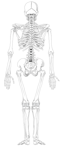
Skeleton diagram no labels
Leg Bone Diagram Labeled / Pin By Pen Duick On ... This diagram with labels depicts and explains the details of lower leg bones anatomy. Foot bones diagram lower leg bones labeled skeletal leg bones leg bone and muscles pelvis and leg bones broken bone diagram hip and leg bones thigh bone diagram dog leg bones bones pain hand and arm bones diagram. Anatomy of the spine and back - e-Anatomy - IMAIOS On "Anatomical parts" the user can choose to display the various structures in colored illustrations of the anatomy of the back and spine: vertebrae, bones, joints, ligaments, muscles, muscular system, fascia, arteries, veins, nerves and various adjacent organs. Diagram of costovertebral joints anatomy (A. Micheau, MD , E-anatomy , Imaios) Clavicle - Definition, Location, Anatomy, & Labeled Diagram Clavicle, commonly known as collarbone, is a slender, S-shaped, modified long bone located at the base of the neck. It is the only long bone of the body that lies horizontally. The term clavicle comes from the Latin word ' clavicula ', meaning 'little key', as its shape resembles an old-fashioned key. Also, the bone rotates along its ...
Skeleton diagram no labels. Chicken Skeleton Anatomy with Labeled Diagram ... This article will show you the detailed anatomy of all bones from the chicken skeleton with a labeled diagram. I will also enlist the peculiar anatomical features of every bone of chicken or bird with their short description. This might be a great resource if you want to know the details on chicken leg bone anatomy, chicken wing bone, and skull. Free Skeletal System Worksheets and Printables Label the Skeleton Activity - This worksheet walks your children through labeling a skeleton. You'll label the main bones of the body. Be sure to scroll down and you'll find worksheets to learn the cranial bones along with the facial bones. Printable Label a Skeleton Worksheets - These skeleton worksheets are a great addition to your lesson plans. Horse Skeleton Anatomy - Osteological Features of Bones ... Horse skeleton anatomy diagram Few special osteological features from the axial and appendicular skeleton of a horse - The skull of a horse is long and four-sided. You will find an extensive foramen lacerum in the horse skull. There is no cornual process in horse skull. The fusion between the two haves of the mandible is complete. Human Skeleton Diagram Blank - free printable skeleton ... Human Skeleton Diagram Blank - 16 images - printable human skeleton diagram graphic by, exercise 12 the fetal skeleton flashcards easy notecards, muscle diagram anatomy system human body anatomy, define the following views and points of axial skeleton in,
Female Body Diagram: Parts of a Vagina, Location, Function Vagina: The vagina is a muscular canal that connects the cervix and the uterus, leading to the outside of the body. Parts of the vagina are rich in collagen and elastin, which give it the ability to expand during sexual stimulation and childbirth. Cervix: The cervix is the lower part of the uterus that separates the lower uterus and the vagina and may play a role in lubrication. Labeled atlas of anatomy: illustrations of the dog - IMAIOS Anatomy of the dog - Illustrated atlas. This modules of vet-Anatomy provides a basic foundation in animal anatomy for students of veterinary medicine. This veterinary anatomical atlas includes selected labeling structures to help student to understand and discover animal anatomy (skeleton, bones, muscles, joints, viscera, respiratory system ... CGAL 5.4 - Manual: Package Overview The 2D Voronoi diagram adaptor package provides an adaptor that adapts a 2-dimensional triangulated Delaunay graph to the corresponding Voronoi diagram, represented as a doubly connected edge list (DCEL) data structure. The adaptor has the ability to automatically eliminate, in a consistent manner, degenerate features of the Voronoi diagram, that are artifacts of the … Skin Diagram Without Labels - connective tissue anatomy ... Skin Diagram Without Labels. Here are a number of highest rated Skin Diagram Without Labels pictures upon internet. We identified it from reliable source. Its submitted by government in the best field. We take this kind of Skin Diagram Without Labels graphic could possibly be the most trending topic gone we part it in google help or facebook.
Anatomical Line Drawings - Medscape Surface Anatomy - lateral views - male. go to drawing without labels. Surface Anatomy - lateral views - female. go to drawing without labels. Surface Anatomy - Child - anterior view & posterior ... Dog Anatomy from Head to Tail - dummies Head's up on dog parts Starting from the head, a dog is made up of the Nose: Dog noses are often cold and wet, and of course, they usually get stuck where they're not wanted. The muzzle (foreface) comprised of the upper and lower jaws.. The stop is an indentation (sometimes nonexistent) between the muzzle and the braincase or forehead.. The forehead (braincase) is the portion of the head ... Anatomy of The Human Ribs - With Full ... - Dislocated Rib The Anatomy of the Human Ribs (costae) are one of the integral parts of the chest wall; they make up the lateral part of our body, its anterior and posterior wall and they entirely build the lateral parts of the chest wall.. The anatomy of the human ribs is made up of 24 ribs. These ribs are parted in 12 pairs (each on the left and right side of the chest wall), with the sternum, metasternum ... Learn all muscles with quizzes and labeled diagrams | Kenhub Labeled diagram View the muscles of the upper and lower extremity in the diagrams below. Use the location, shape and surrounding structures to help you memorize each muscle. Once you're feeling confident, it's time to test yourself. Unlabeled diagram See if you can label the muscles yourself on the worksheet available for download below.
Human skeleton labelling sheetsHuman Skeleton
File:Diagram of the human heart (cropped).svg - Wikipedia No labels version العربية ܐܪܡܝܐ azərbaycanca bosanski ... Add cardiac skeleton. Inferior vena cava more wide. Add aorta in bottom. Add source veins of superior vena cava. Brachiocephalic trunk more wide and separated. Added shadows. Left main pulmonary artery with its first division. 07:02, 2 June 2006: 650 × 650 (26 KB) Yaddah: Diagram of the human heart, created by …
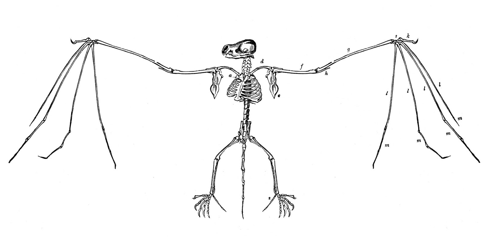
Vintage Halloween Clip Art - Super Creepy Bat Skeletons - The Graphics Fairy
Cladogram- definition, features, parts, examples (vs ... Cladogram Definition. A cladogram is the graphical representation of the hypothetical relationship (phylogenetic relationship) between different groups of organisms. It is used in the phylogenetic analysis of organisms to determine the evolutionary relationship between them. The cladogram is derived from Greek words clados and gramma where 'clados' means branch and 'gramma' means ...
Biological Anthropology/Unit 2: Non-human Primates/Primate Skeletal Anatomy - WikiEducator
FREE Human Body Systems Labeling with Answer Sheets The free skeletal system labeling sheet includes a fill-in-the-blanks labeling of the main bones on the body. The free respiratory system labeling sheet includes a blank diagram to fill in the trachea, bronchi, lungs, and larynx. The free nervous system labeling sheet includes blanks to label parts of the brain, spinal cord, ganglion, and nerves.

Skeleton Labeling Page | Homeschool Science | Pinterest | Skeletons, Homeschool and Body systems
Labeled imaging anatomy cases | Radiology Reference ... URL of Article. This article lists a series of labeled imaging anatomy cases by body region and modality. On this page: Article: Brain. Head and neck. Spine. Chest. Abdomen and pelvis.

Appendicular Skeleton Labeling Blank | Skeletal System Diagram No Labels | Homeschool ...
Anatomy Project Neck. · Connecting the shaft and head of the femur. · Projects superior and medial from the shaft to the head. · In addition to projecting superior and medial from the shaft of the femur, the neck also projects somewhat anterior. · The amount of forward projection is extremely variable, but on an average is from 12° to 14°.

Human Skeleton Blank Clip Art at Clker.com - vector clip art online, royalty free & public domain
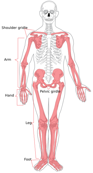
File:Human skeleton front en.svg - Wikipedia English: diagram of a human female skeleton. the Red lines point individual bones and the names are writen in singular, the blue lines connect to group of bones and are in plural form. Date: 3 January 2007: Source: Own work. Image renamed from File:Human skeleton front.svg: Author: LadyofHats Mariana Ruiz Villarreal: Permission (Reusing this file) Public domain Public …

Skeleton Posterior Clip Art at Clker.com - vector clip art online, royalty free & public domain
Human Skeleton Diagram Unlabeled - anatomy of a skeleton ... Human Skeleton Diagram Unlabeled - 15 images - primary years 5 6 news tns administered by mr c, urinary system diagram medical art library, human skeleton labeled, skeletal system creationwiki the encyclopedia of,
FREE! - Human Skeleton Labelling Sheet (teacher made)
Diagram of Human Heart and Blood Circulation in It | New ... Four Chambers of the Heart and Blood Circulation. The shape of the human heart is like an upside-down pear, weighing between 7-15 ounces, and is little larger than the size of the fist. It is located between the lungs, in the middle of the chest, behind and slightly to the left of the breast bone. The heart, one of the most significant organs ...

Claye Willcox Athlete Dev.: Muscular/Skeletal Systems + Joints
To Blank Label Skeleton [XRG57Z] Blank Skeleton Diagram to Label. Using the mouse, color the individual parts of the. 14) are also known as the solar panels. LABELING EXERCISE: BONES OF THE AXIAL AND APPENDICULAR SKELETON. Save your finished labeled picture. Appendicular Skeleton - upper limb. 260 Labels Per Sheet.

Skeleton Labeling Page | Homeschool Science | Pinterest | Skeletons, Homeschool and Body systems
How to Create a Fishbone Diagram for Medical Diagnosis ... A fishbone diagram, also known as an Ishikawa diagram or a cause-and-effect diagram, is a model used to identify potential root causes of a problem or an outcome. In health care, using a fishbone diagram to simplify medical diagnoses can help patients gain a better understanding of their medical conditions.
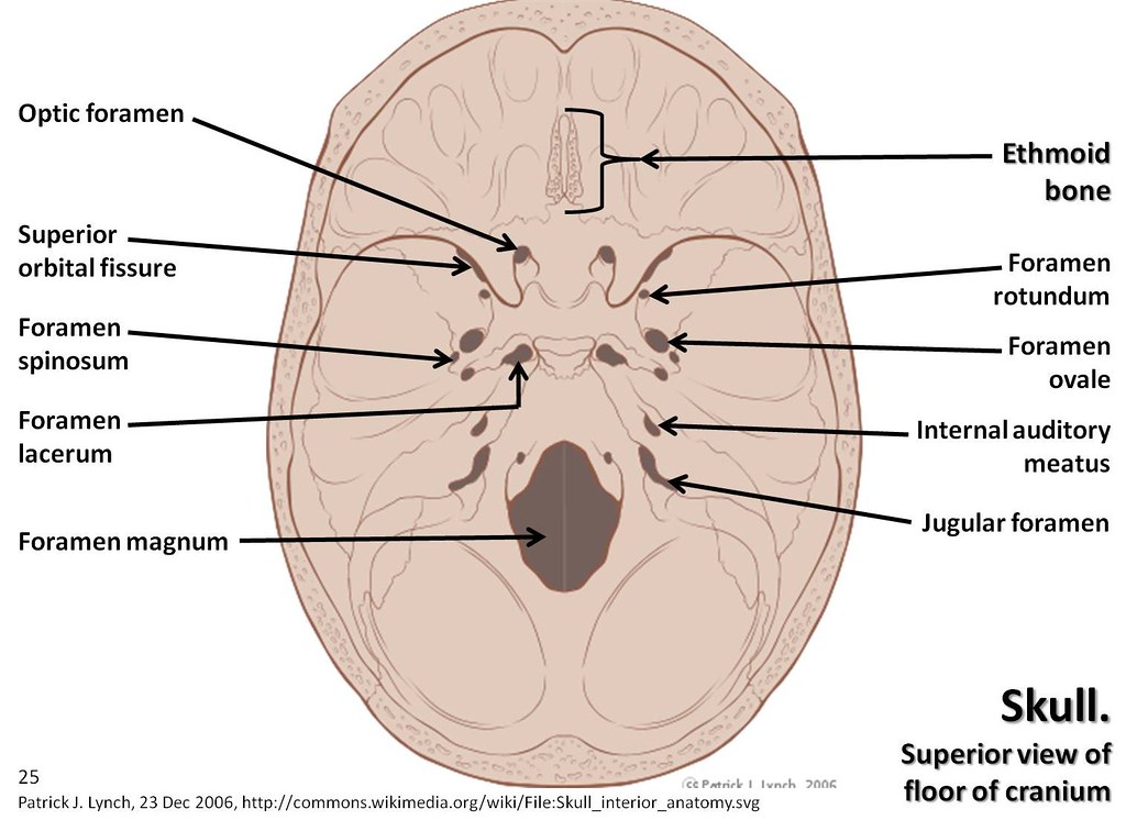
Skull diagram, superior view of floor of cranium with labe… | Flickr
Scapula - Parts, Anatomy, Location, Functions, & Labeled ... What is Scapula. The scapula, alternatively known as the shoulder blade, is a thin, flat, roughly triangular-shaped bone placed on either side of the upper back. This bone, along with the clavicle and the manubrium of the sternum, composes the pectoral (shoulder) girdle, connecting the upper limb of the appendicular skeleton to the axial skeleton.
Appendicular Skeleton Diagram Clip Art at Clker.com - vector clip art online, royalty free ...
How to identify mammal skulls | BBC ... - Discover Wildlife Deer skulls have no upper incisors; the cheek teeth are all very similar and designed for grinding. Male deer skulls are easily recognised by antlers; if antlers are not present, the short, upwardly directed pedicel is cut flat and points backward. With sheep, the horn boss, or boney growth, is pointed and tapered and curves backward and downward.
Human Skeleton Back No Text No Color Clip Art at Clker.com - vector clip art online, royalty ...
Bones of Contention Quiz | Human Body | 15 Questions 2 parietal bones (the sides and top - joining with the frontal) 1 sphenoid bone (front middle of skull) 2 temporal bones (the sides and base) Mandible The other part of the skull is made of your facial bones, which (amongst other things) is what gives your face its distinct shape. Of the 14 facial bones, the mandible, or jaw bone, is the largest.
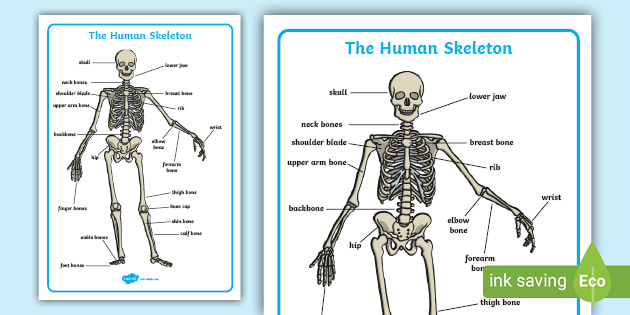
FREE! - Human Skeleton Labelling Sheet | Label the Skeleton Activity
Clavicle - Definition, Location, Anatomy, & Labeled Diagram Clavicle, commonly known as collarbone, is a slender, S-shaped, modified long bone located at the base of the neck. It is the only long bone of the body that lies horizontally. The term clavicle comes from the Latin word ' clavicula ', meaning 'little key', as its shape resembles an old-fashioned key. Also, the bone rotates along its ...
KS2 Label the skeleton - Teaching resources
Anatomy of the spine and back - e-Anatomy - IMAIOS On "Anatomical parts" the user can choose to display the various structures in colored illustrations of the anatomy of the back and spine: vertebrae, bones, joints, ligaments, muscles, muscular system, fascia, arteries, veins, nerves and various adjacent organs. Diagram of costovertebral joints anatomy (A. Micheau, MD , E-anatomy , Imaios)

Skeleton Labels | SEN Teacher | Pinterest | Skeleton labeled, Science resources and Teacher
Leg Bone Diagram Labeled / Pin By Pen Duick On ... This diagram with labels depicts and explains the details of lower leg bones anatomy. Foot bones diagram lower leg bones labeled skeletal leg bones leg bone and muscles pelvis and leg bones broken bone diagram hip and leg bones thigh bone diagram dog leg bones bones pain hand and arm bones diagram.
Human Skeleton Diagram Without Labels - ClipArt Best
Post a Comment for "41 skeleton diagram no labels"