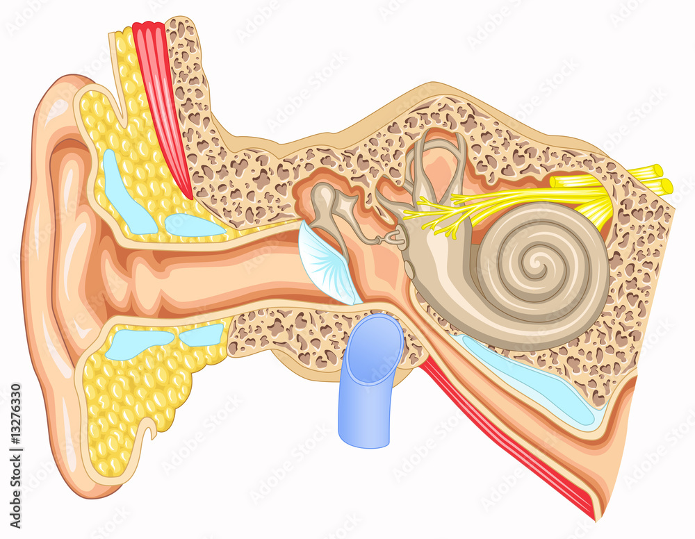
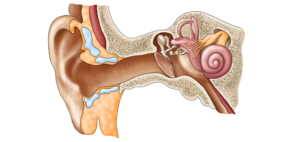
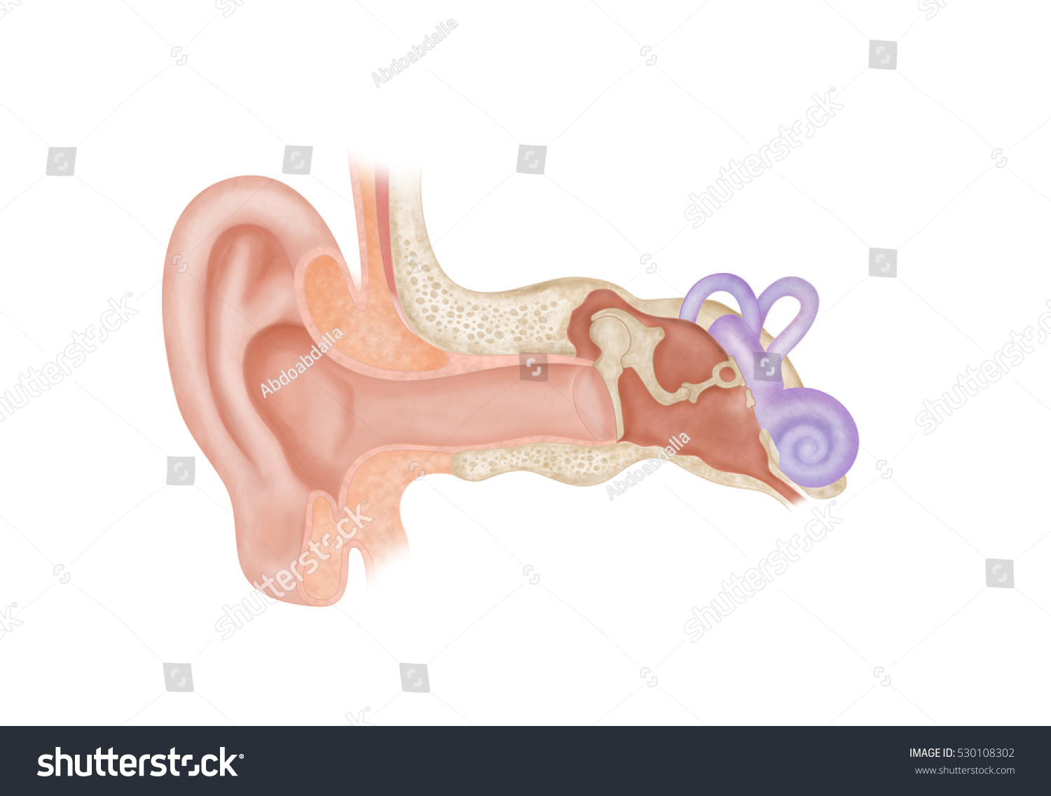
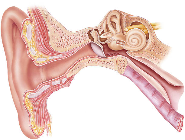
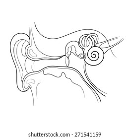
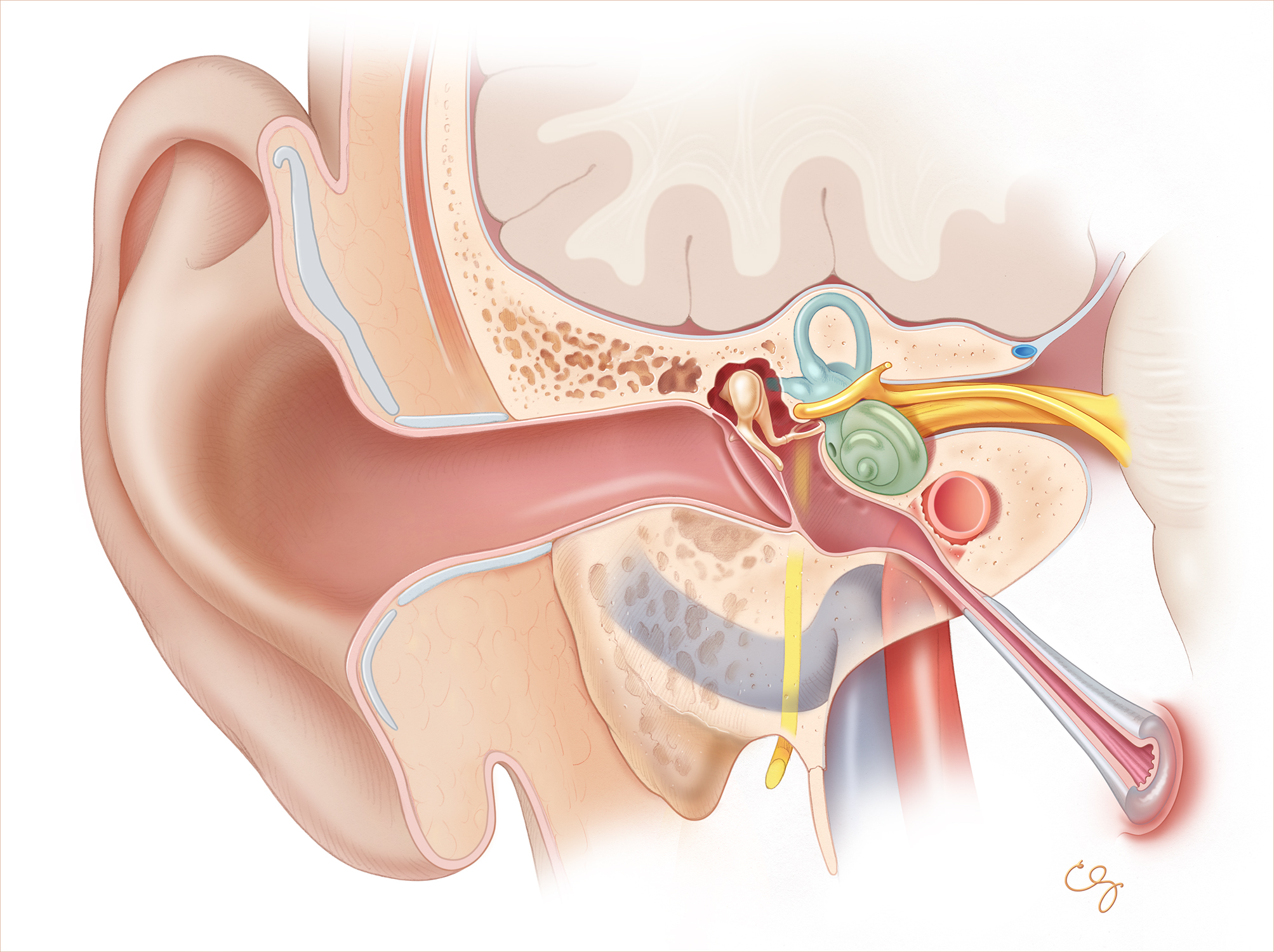
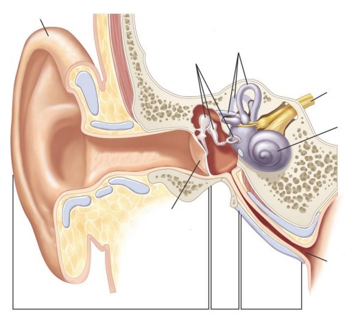
40 ear anatomy without labels
Normal chest MDCT with anatomic labels | e-Anatomy - e-Anatomy … 10/03/2022 · Normal anatomy of the thorax on labeled Chest CT: radiological anatomy of the lungs, mediastinal lymph nodes, trachea, bronchi, pleural cavity, heart and pulmonary vessels. Anatomy of the Ear | Geeky Medics The tympanic membrane, or eardrum, marks the border between the external and middle ear. It is formed of a middle layer of connective tissue with a layer of skin on its lateral surface (facing the external acoustic meatus) and mucous membrane on its medial surface (facing the middle ear).
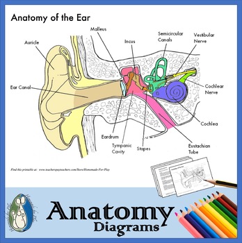
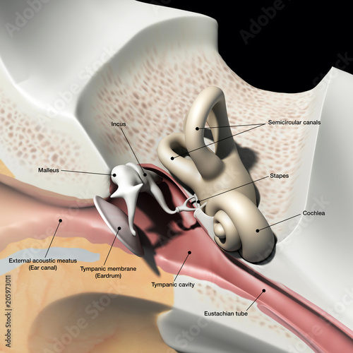
Human Ear: Structure and Anatomy - Online Biology Notes Ear ossicles: The three ear ossicles (malleus, incus and stapes) form a chain of lever extending from tympanic membrane to inner ear. The ear ossicles transmit sound wave from ear drum to inner ear. Ear ossicles communicate the ear drum with internal ear through fenestra ovalis ( oval window). The ear ossicles are;
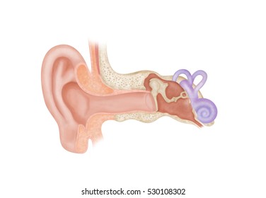
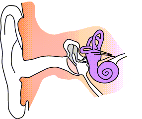
Ear anatomy without labels
Picture of the Ear: Ear Conditions and Treatments - WebMD Earache: Pain in the ear can have many causes. Some of these are serious, some are not serious. Otitis media (middle ear inflammation): Inflammation or infection of the middle ear (behind the ... Anatomy Lecture Midterm Flashcards | Quizlet Study with Quizlet and memorize flashcards containing terms like Place a single word into each sentence to make it correct. Then rearrange the sentences into the correct order to explain the process of the cardiocyte action potential. +30mV Chloride Ion Negative Positive -55mV Resting Calcium ion Cardiomyocyte Once these channels close, potassium ions flow out quickly and … › enAnatomy, medical imaging and e-learning for healthcare ... IMAIOS and selected third parties, use cookies or similar technologies, in particular for audience measurement. Cookies allow us to analyze and store information such as the characteristics of your device as well as certain personal data (e.g., IP addresses, navigation, usage or geolocation data, unique identifiers).
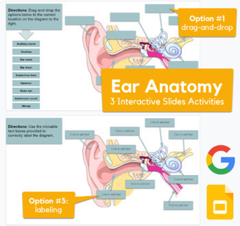
Ear anatomy without labels. Image result for ear structure without label | Ear diagram, Human ear ... Feb 12, 2018 - Image result for ear structure without label. Feb 12, 2018 - Image result for ear structure without label. Pinterest. Today. Explore. When the auto-complete results are available, use the up and down arrows to review and Enter to select. Touch device users can explore by touch or with swipe gestures. Blank ear diagrams and quizzes: The fastest way to learn ear anatomy Ear diagrams (labeled and unlabeled) Accelerate your learning with interactive quizzes Sources + Show all Ear anatomy overview Although it's not obvious to look at, the ear is anatomically divided into three portions: External (outer) ear Middle ear Inner ear As you can imagine, there's a lot of associated anatomy to learn for each portion! Ear Diagram and Labeling Worksheet / Worksheet - Twinkl This allows you to tailor the task to the individual abilities of your learners. The first worksheet presents an ear with annotations showing the first letters of its key features. For example, a label marked 'P' links to the Pinna (outer ear). The second page shows an ear diagram without labels. The final page shows the labels linking to the ... Enchanted Learning Moved Permanently. The document has moved here.
Image result for ear structure without label | Ear anatomy, Human ear ... Each nephron is made of two main parts called malpighian body and covoluted tubule.Malphigian body is double layered cup also called bowman`s capsule. The inner cup consists network of capillaries called Glomerulus. Now let`s start the diagram. 1.Draw a egg shape and make a folded curve as shown in the figure and… L Amy Yee Anatomy Ear Anatomy - Worksheet Activity - Ask A Biologist Ear Anatomy Activity. The parts of a ear have been labeled. Your challenge is to write the correct name for each part. To learn more, visit. Anatomy of the eye: Quizzes and diagrams | Kenhub How to learn the parts of the eye. Found within two cavities in the skull known as the orbits, the eyes are surrounded by several supporting structures including muscles, vessels, and nerves. There are 7 bones of the orbit, two groups of muscles (intrinsic ocular and extraocular), three layers to the eyeball … and that's just the beginning. Human Ear Diagram - Bodytomy Auditory Ossicles: The three small bones in the middle ear, called malleus, stapes, and incus, are connected. These bones together are called the auditory ossicles, and their purpose is to let the sound that strikes the eardrum, further into the inner ear.
Anatomy of the Ear | Inner Ear | Middle Ear | Outer Ear The Outer Ear The outer ear includes: auricle (cartilage covered by skin placed on opposite sides of the head) auditory canal (also called the ear canal) eardrum outer layer (also called the tympanic membrane) The outer part of the ear collects sound. Sound travels through the auricle and the auditory canal, a short tube that ends at the eardrum. Ear Anatomy without Labels, Digital Art - Shutterstock Ear Anatomy Without Labels Digital Art Stock Illustration 530108302 Download for free See more Popularity score High Usage score High usage Superstar Shutterstock customers love this asset! Item ID: 530108302 Ear Anatomy without Labels, Digital Art Formats 8976 × 6201 pixels • 29.9 × 20.7 in • DPI 300 • JPG Anatomy, medical imaging and e-learning for healthcare IMAIOS and selected third parties, use cookies or similar technologies, in particular for audience measurement. Cookies allow us to analyze and store information such as the characteristics of your device as well as certain personal data (e.g., IP addresses, navigation, usage or geolocation data, unique identifiers). Human Middle Ear Anatomy Cross Section View With Labels Stock Photo ... Description Computer generated image of the human middle ear bones and inner ear with anatomical labeling. 1 credit Essentials collection for this image $4 with a 1-month subscription (10 Essentials images for $40) Continue with purchase View plans and pricing Includes our standard license. Add an extended license. Credit: Hank Grebe
Well-Labelled Diagram Of Ear With Explanation - BYJUS Eustachian Tube is a tube that connects the middle ear to the back of the nose. It helps to maintain equal pressure in the middle ear which facilitates the proper transmission of sound waves. The Inner ear consists of Cochlea that comprises the nerves of hearing. Semicircular canals contain the receptors that help in maintaining balance.
14,026 Human ear anatomy Images, Stock Photos & Vectors - Shutterstock 14,026 human ear anatomy stock photos, vectors, and illustrations are available royalty-free. See human ear anatomy stock video clips Image type Orientation Color People Artists Sort by Popular Healthcare and Medical Anatomy ear cochlea medicine inner ear organ human body biology diagram Next of 141
The Ear: Anatomy, Function, and Treatment - Verywell Health The middle ear (also known as the tympanum or tympanic cavity) is a complicated network of tunnels, chambers, openings, and canals mostly inside openings within the temporal bone on each side of the skull. The 2 largest chambers are called the middle ear space and mastoid.
Inner Ear Anatomy, Function, and Health Inner ear function. The inner ear has two main functions. It helps you hear and keep your balance. The parts of the inner ear are attached but work separately to do each job. The cochlea works ...
Ear (Anatomy): Overview, Parts and Functions | Biology Dictionary The ear canal is the opening through which sound waves enter the middle ear. It serves to further focus and concentrate the vibrations collected by the pinna, ensuring that the vibrations will be clear and strong enough to be amplified and turned into nerve impulses. The ear canal is only 2-3 centimeters deep - a little bit less than one inch.
quizlet.com › 414265947 › anatomy-lecture-midtermAnatomy Lecture Midterm Flashcards | Quizlet Study with Quizlet and memorize flashcards containing terms like Place a single word into each sentence to make it correct. Then rearrange the sentences into the correct order to explain the process of the cardiocyte action potential. +30mV Chloride Ion Negative Positive -55mV Resting Calcium ion Cardiomyocyte Once these channels close, potassium ions flow out quickly and restore the ...
human ear | Structure, Function, & Parts | Britannica human ear, organ of hearing and equilibrium that detects and analyzes sound by transduction (or the conversion of sound waves into electrochemical impulses) and maintains the sense of balance (equilibrium). The human ear, like that of other mammals, contains sense organs that serve two quite different functions: that of hearing and that of postural equilibrium and coordination of head and eye ...
Outer Ear: Anatomy, Location, and Function - Verywell Health Fossa, superior crus, inferior crus, and antihelix: These sections make up the middle ridges and depressions of the outer ear. The superior crus is the first ridge that emerges moving in from the helix. The inferior crus is an extension of the superior crus, branching off toward the head. The antihelix is the lowest extension of this ridge.
Anatomy and Physiology of the Ear - Stanford Children's Health External auditory canal or tube. This is the tube that connects the outer ear to the inside or middle ear. Tympanic membrane (eardrum). The tympanic membrane ...
Human Ear Anatomy - Parts of Ear Structure, Diagram and Ear Problems The external (outer) ear consists of the auricle, external auditory canal, and eardrum (Figure 1 and 2). The auricle or pinna is a flap of elastic cartilage shaped like the flared end of a trumpet and covered by skin. The rim of the auricle is the helix; the inferior portion is the lobule. Ligaments and muscles attach the auricle to the head.
Label Parts of the Human Ear - University of Dayton Parts of the Ear. Select the correct label for each part of the ear. Click on the Score button to see how you did. Incorrect answers will be marked in red.
Amazon.com: Flents Foam Ear Plugs, 10 Pair with Case for … If you have average ear canals and want the most noise blocking ear plugs, I would recommend these.Light Green Flents Protech Contour:I like contour ear plugs because my ears are sensitive. These are pretty large ear plugs and not the best for those with small ears. They were the best price of all the earplugs I tried. If you have average-larger ear canals, are a side sleeper, or …
› 40600784 › LIBRO_PARA_COLOREAR_NETTER(PDF) LIBRO PARA COLOREAR NETTER - Academia.edu Background: The aim of our study was to examine the effect of mild maternal hypothyroidism on the apoptosis of the oocytes in the ovaries of rats in the early postnatal period during formation of oocytes and follicles.
en.wikipedia.org › wiki › Human_penisHuman penis - Wikipedia The distal section of the urethra allows a human male to direct the stream of urine by holding the penis. This flexibility allows the male to choose the posture in which to urinate. In cultures where more than a minimum of clothing is worn, the penis allows the male to urinate while standing without removing much of the clothing.
"Outer Ear Anatomy Neutral Pattern (without center label)" T-shirt for ... Buy "Outer Ear Anatomy Neutral Pattern (without center label)" by StacyAnnDesigns as a Essential T-Shirt.
Ear Anatomy - Outer Ear | McGovern Medical School The medical term for the outer ear is the auricle or pinna. The outer ear is made up of cartilage and skin. There are three different parts to the outer ear; the tragus, helix and the lobule. EAR CANAL The ear canal starts at the outer ear and ends at the ear drum. The canal is approximately an inch in length.
› en › e-AnatomyNormal chest MDCT with anatomic labels | e-Anatomy - IMAIOS Mar 10, 2022 · Normal anatomy of the thorax on labeled Chest CT: radiological anatomy of the lungs, mediastinal lymph nodes, trachea, bronchi, pleural cavity, heart and pulmonary vessels. × Your email address is not verified.
Ear Anatomy Images | McGovern Medical School The malleus is the middle ear bone which is attached to the drum and easily identified. The middle ear space can be seen through the ear drum and a portion of the incus (another middle ear bone) can be identified. Normal Left Ear Drum This is a left ear drum of a ten year old. The drum looks healthy and has a nice gray color to it.
Human Body Parts Images Without Labels - Free Vector Download 2020 Human ear diagram with labels and label of anatomy labeling the ear purposegames nose diagram with label diagrams all labels human ear the ear diagram without labels anatomy human charts. Illustration Of Body Parts Labels It is certainly the most widely studied structure the world over. Human body parts images without labels. Download body ...
Outer Ear Anatomy Colorful Pattern (without center label) Tote Bag Buy "Outer Ear Anatomy Colorful Pattern (without center label)" by StacyAnnDesigns as a Tote Bag.
› Flents-Quiet-Contour-Plugs-PairAmazon.com: Flents Foam Ear Plugs, 10 Pair with Case for ... Specialized contour shape creates a snug and comfortable fit in your ear 20 PACK - Product comes in a convenient pack of 10 pair NOISE REDUCTION RATING - Ear plugs rated for 33 decibels SAFE TO USE - Product is safe to use and designed to keep your inner ear safe when inserted
Image result for ear structure without label - Pinterest Preschool Kid Learning Ear Coloring Pages to Color, Print and Download for Free along with bunch of favorite Ear coloring page for kids. Simply do online coloring for Preschool Kid Learning Ear Coloring Pages directly from your gadget, support for iPad, android tab or using our web feature. D. Darina Lețu. Doodle and Art.
Ear Labels Flashcards | Quizlet Terms in this set (16) auricle external auditory canal tympanic membrane malleus (hammer) Incus (anvil) stapes (stirrup) auditory/eustachian/pharyngotympanic tube vestibules semicicular canals Ampulla of semicircular canals round window oval window and round window cochlea snail cochlear duct in cochlea vestibular nerve
255 Human Ear Diagram Premium High Res Photos - Getty Images external auditory canal of human ear (with labels). - human ear diagram stock illustrations anatomy of the ear and nose engraving antique illustration, published 1851 - human ear diagram stock illustrations
apps.apple.com › us › appThe Human Body by Tinybop 4+ - App Store + Feed the body, make it run and breathe, assemble and pull apart a skeleton, see how the eye sees, watch sound vibrations travel through the ear canal, and more. + Learn new vocabulary with text labels in 50+ languages. + Create a dashboard to change languages, delete accounts, and support your kids’ learning.
(PDF) LIBRO PARA COLOREAR NETTER - Academia.edu Gray - Anatomy of the Human Body. ALina ivanovic. Download Free PDF View PDF. Human Anatomy. Stella Pl. Download Free PDF View PDF. anatdescsurgi grayrich.pdf. Anis Zamir. Download Free PDF View PDF. Apoptosis of the oocytes of hypothyroid neonatal rats. Neda Drndarevic. Background: The aim of our study was to examine the effect of mild maternal …
label the ear worksheet 14 Best Images Of Ear Hearing Worksheets - Listening Ear Craft Template ear worksheet diagram inner anatomy answers parts eye animals physiology labelled worksheets ossicles senses auditory middle hearing outer pinna canal Ear Diagram Without Labels & With Them - Labelling Worksheet
Ear Diagram Without Label Vector Free | AI, SVG and EPS Free Ear Diagram Without Label vector download in AI, SVG, EPS and CDR. Browse our Ear Diagram Without Label images, graphics, and designs from +79.322 free ...
› enAnatomy, medical imaging and e-learning for healthcare ... IMAIOS and selected third parties, use cookies or similar technologies, in particular for audience measurement. Cookies allow us to analyze and store information such as the characteristics of your device as well as certain personal data (e.g., IP addresses, navigation, usage or geolocation data, unique identifiers).
Anatomy Lecture Midterm Flashcards | Quizlet Study with Quizlet and memorize flashcards containing terms like Place a single word into each sentence to make it correct. Then rearrange the sentences into the correct order to explain the process of the cardiocyte action potential. +30mV Chloride Ion Negative Positive -55mV Resting Calcium ion Cardiomyocyte Once these channels close, potassium ions flow out quickly and …
Picture of the Ear: Ear Conditions and Treatments - WebMD Earache: Pain in the ear can have many causes. Some of these are serious, some are not serious. Otitis media (middle ear inflammation): Inflammation or infection of the middle ear (behind the ...












Post a Comment for "40 ear anatomy without labels"